Introduction to MRI
This handout describes different ways in which NMR spectrometers can work, and the basics of MRI as a medical diagnostic tool. As a painless, noninvasive imaging technique with little in the way of risks, MRI will become even more popular with time. Since the principles underlying MRI are the same as those behind NMR, this is an appropriate time to learn the answer to the question "How does MRI work?".
1.0 Different ways to carry out the NMR experiment.
1.1 Fixed magnetic field, scan radio frequencies. This is conceptually the simplest NMR experiment, in which the sample is placed in a homogeneous external magnetic field and radio frequencies are scanned.
1.1A Every time a frequency matches the resonant frequency of a particular type of proton in a sample, the energy is absorbed and the NMR spectrometer records this absorption as a peak that is plotted at the appropriate ppm with the appropriate peak height and splitting pattern, etc.
1.1B Recall that using ppm as the frequency scale insures that magnetic field strength differences between different machines are scaled out of the spectra so that direct comparisons can be made.
1.2 Fixed radio frequency, scan magnetic field strength. Since the resonant frequency (i.e. energy difference between +1/2 and –1/2 spin states) of a given proton is proportional to the external magnetic field strength, scanning magnetic field strength in the presence of a fixed radio frequency irradiation is an alternative way to generate an NMR spectrum.
1.2A A peak is recorded when the scanned magnetic field reaches exactly that needed for a given proton to resonate at the fixed radio frequency being used. The NMR spectrometer must then convert this information to the frequency vs. absorbance plot. In other words, even though magnetic field is scanned, not frequency, in spectrometers of this type, NMR spectra are always converted to a form as if frequency were scanned in a fixed magnetic field so that spectra recorded on different machines can be easily compared.
1.2B It turns out that for technical reasons, it is actually better to make this kind of fixed radio frequency irradiation, scanning magnetic field spectrometer than one with a fixed magnetic field and scanning radio frequency irradiation.1.3 Fixed magnetic field, irradiate with all radio frequencies simultaneously (“broad band irradiation”) to flip all spins instantaneously, monitor the spins flipping back to lower energy state (-1/2) to deduce resonant frequencies.
1.3A You will have to take it on faith that the rate at which irradiated nuclear spins "relax" back from the –1/2 spin state to the lower energy +1/2 spin state is directly related to their resonant frequency in the same external magnetic field. Thus, the observed “relaxation rates” can be converted to absorbance frequencies (ppm) through mathematical manipulation of the data.
1.3B The mathematical treatment utilizes an algorithm called Fourier Transform (FT), so this type of spectrometer is called an FT-NMR. In simple terms, Fourier Transform takes the time information from the relaxation rates and breaks it up into its component frequency information, which is plotted as peaks on a ppm scale.
1.3C Virtually all new NMR machines are FT-NMR. The huge advantage is that the experiment is extremely fast and can be easily repeated hundreds or thousands of times on the same sample to generate a spectrum with exceptional signal-to-noise even from very dilute samples. In addition, by manipulating exactly how the sample is excited then allowed to relax using so-called pulse sequences, multi-dimensional spectra can be obtained that have phenomenal mounts of information in them, so-called 2D or even 3D NMR (beyond the scope of this class but really cool!)
Bottom line. There are at least three ways that have been used to measure nuclear spin flipping. However, no matter what the method used, the data is always plotted as proton resonance frequency (ppm) that gives chemical shifts and splitting patterns, and for 1H NMR, the area of the peaks is a relative measure of the number of equivalent protons in each set.
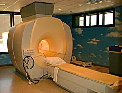
2.1 In MRI, a patient is placed in a very strong magnetic field. The magnet is the large cylinder the patient is moved into, such as that shown below.
2.1A The large magnet aligns the nuclear spins inside the patient, usually protons, in their +1/2 and –1/2 spin states.
2.1B Humans are about 60% water, and water obviously has a large number of protons per unit volume. Fat, containing largely –CH2- groups, also contains a large number of protons per unit volume. Thus, water and fat give relatively strong signals in 1H MRI images.
2.1B Different tissues, including tumors, have different quantities of water/fat, so they show up differently in an MRI image. More protons per unit volume give a larger signal in an image, and vice versa.
2.1C Radio frequency electromagnetic radiation is used to flip the aligned proton nuclear spins.
2.2 The three-dimensional
image of a patient’s body is reconstructed from slices, stacked up like
a stack of CD’s.
2.2A Each slice is created by taking measurements of individual lines through the body, with line measurements taken around a circle on the outside of the patient. These individual measurements are then reconstituted into a slice.
2.2B Many slices are taken and stacked together to give a 3-D map of the interior of a body.
2.3 For a variety of technical reasons, it is the FT-NMR approach used
in MRI machines. This provides for very quick acquisition of the individual
data lines, so that slice and image measurement can be acquired in a few minutes.
It is difficult to overstate the level of mathematical analysis and sophistication
needed to generate a 3D image from the raw individual lines of data gathered.
As you could imagine, the physical basis for MRI is difficult to explain to patients. Nevertheless, it is very important to me that you understand it, so you will be tested on the basics of how NMR and MRI work, along with simply solving chemcial NMR spectra. Below is copied a typical web description of MRI imaging, intended to inform patients. Now that you know the whole story, you can appreciate the details that are missing.
‘Diagnostic Imaging'
Presbyterian Hospital offers a wide variety of services for diagnosis and treatment, many available on an outpatient basis. When you come in for testing or rehab, our skilled staff uses the latest equipment, with an emphasis on your comfort.
Magnetic Resonance Imaging (MRI)
Magnetic Resonance Imaging uses a magnetic field and radio waves to produce a highly accurate view of the inside of any portion of your body without using X-rays. It's painless and extremely safe because no radiation is used. It has no known side effects.
MRI offers a non-invasive way to obtain information about your body. It can lead to early detection and treatment of disease because it makes it possible to see certain types of tissue. It can provide important information about the brain, spine, joints, and internal organs.
Below are a number of websites with useful information about MRI, in the form of detailed tutorials
http://www.cis.rit.edu/htbooks/mri/
http://www.mritutor.org/mritutor/.
Below are links to many different MRI images. Note how each is of a single slice through a particularly informative area of a patient's body.
arm,
In 2003, the Nobel prize in medicine went Paul Lauterbur and Peter Mansfield for their work to help develop MRI.
http://www.nobel.se/medicine/laureates/2003/press.html